Vinmec doctors' breathtaking "race against time" to save the life of a 20-week-old fetus.
To date, more than 30 fetal blood transfusions have been performed at the Vinmec Fetal Medicine Department. The success rate is over 95% – this "telling" figure reflects the high level of expertise in handling high-risk pregnancies that previously offered almost no chance of success.
A challenging pregnancy
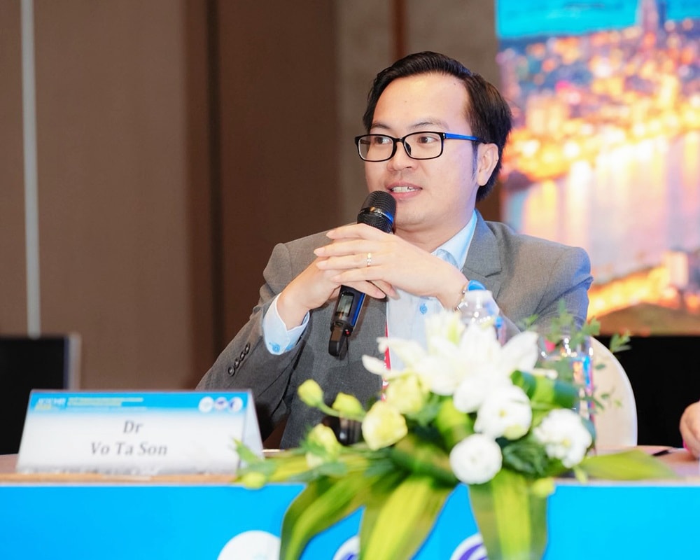
In late November 2025, a pregnant woman at 20 weeks of gestation was transferred to Vinmec Times City Hospital (Hanoi) in a critical condition with fetal hydrops fetalis. Dr. Vo Ta Son, a specialist in fetal medicine at the Women's Health Center, Vinmec Times City International General Hospital, was the doctor who received the patient.Recalling the case, she said, "The fetus had severe anemia, generalized hydrops fetalis, and multiple membrane effusions. These signs indicated a very high risk of the baby not surviving without timely intervention."
Dr. Son and the experts determined that the most likely cause was the pregnant woman's infection with Parvovirus B19 – an agent that can inhibit red blood cell production, leading to severe anemia, heart failure, and fetal hydrops. After thorough consultation and with the family's consent, the team immediately decided to proceed with blood transfusion for the fetus in the womb.
In the field of fetal medicine, this is one of the most challenging intervention techniques, requiring seamless coordination between technology and the doctor's experience. The entire procedure demands extremely high precision; the fetus has a chance of survival if the intervention is performed at the right time, but there is also a potential risk of miscarriage during the procedure.
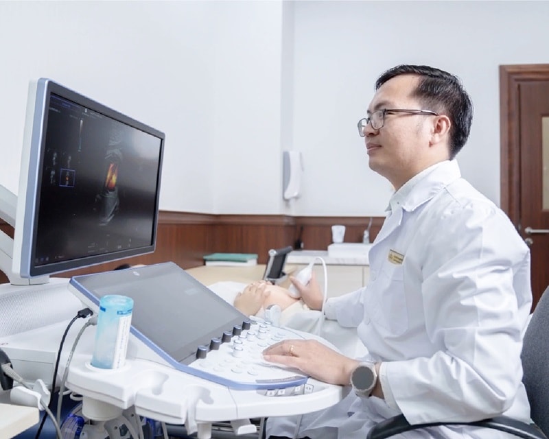
"At 20 weeks of gestation, the fetal umbilical vein is only about 2-3 mm in diameter, roughly the size of a toothpick tip. To access this structure, we have to use a specialized needle to precisely insert into the fetal blood vessel under the guidance of a high-resolution ultrasound system," Dr. Son explained.
Twenty-four hours after the intervention, the indicators showed that the fetus was beginning to respond – a fragile but hopeful sign. However, at the follow-up ultrasound one week later, the situation became more complicated when the doctor discovered that the fetus had relapsed anemia.
Faced with this situation, the team had a difficult decision: Should they proceed with a second intervention or stop? Choosing to continue meant accepting additional risks, but it was also the only chance to save the fetus. After numerous interdisciplinary consultations and thorough consideration, the team decided to proceed with a second blood transfusion.
After the second intervention, the fetus's condition improved significantly, with each indicator gradually stabilizing. After a challenging pregnancy, the baby was born healthy at 39 weeks. The entire family and medical team were overwhelmed with joy witnessing this moment, a seemingly fragile life being saved through extraordinary efforts.
Accompanying the "seed of life": A journey to change the fate of unborn children.
Looking back, Dr. Son shared that his journey into the field of Obstetrics and Gynecology - Fetal Medicine began with a very simple desire: to be present during the first moments of life. However, during his work, he gradually realized that behind each pregnancy there is not only joy but also many challenges related to pathology, especially congenital abnormalities.
"When I was exposed to fetal morphology ultrasound, I realized that with just a small probe, doctors can 'see the future' of a baby, and sometimes we can even change it. That's both miraculous and places a great responsibility," Dr. Son confided.
This realization motivated doctors to pursue the field of prenatal diagnosis and fetal intervention in greater depth, with the hope of not only early detection but also the ability to change the prognosis for the fetus while still in the womb.
Having received thorough training at numerous major medical centers both domestically and internationally, Dr. Son stated that his studies in an international environment have given him a more comprehensive understanding of fetal medicine. Accordingly, diagnosis has moved beyond simply detecting abnormalities, now encompassing the analysis of causes, predictive prognoses, and individualized pregnancy management for each specific case.
In addition to directly participating in complex fetal ultrasound and intervention procedures, Dr. Son is also a researcher in the field of fetal medicine. He regularly presents at national and international professional conferences. Recently, he co-authored a research paper on the early diagnosis of fetal brain development abnormalities, published in the official journal of the International Society of Obstetric and Gynecological Ultrasound – one of the world's leading prestigious journals in this field.
For Dr. Son, scientific research is not just about academic publications, but also about bringing the latest advances into clinical practice. "Each study helps us better understand the nature of the disease and improve each treatment decision, thereby giving fetuses a better chance of survival," he shared.
At the intersection of science and practice, the journey of fetal medicine practitioners is not just about diagnosis, but also about the relentless effort to change prognoses, opening up more fulfilling beginnings for "new life" from within the mother's womb.