Three-dimensional biomolecular imaging technique wins Nobel Prize in Chemistry
Three chemists, Jacques Dubochet, Joachim Frank, and Richard Henderson, won the Nobel Prize for developing a method for creating 3D images of the structures that make up life.
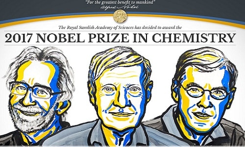 |
| Portraits of the three scientists who won the Nobel Prize in Chemistry this year: Jacques Dubochet, Joachim Frank and Richard Henderson. |
This year’s Nobel Prize in Chemistry honors a group of scientists who invented techniques that allowed them to record the intricate molecular machinery of life in detail, according to the Guardian. The Swedish Academy of Sciences announced the winners today in Stockholm to Jacques Dubochet, Joachim Frank and Richard Henderson. The prize, worth more than $1.1 million, will be split equally between the three.
The technique developed by the team, called cryo-electron microscopy, enables the study of biomolecular structures at high resolution for the first time, a revolutionary achievement in biochemistry.
Before the groundbreaking work, electron microscopes were thought to be suitable only for imaging dead matter, as the powerful electron beams would destroy biological material. Henderson, a Scottish scientist and professor at the MRC Laboratory of Molecular Biology, succeeded in using an electron microscope to create the first three-dimensional image of a protein at atomic resolution. The result helped demonstrate the potential of the technology.
Frank, a German professor at Columbia University in New York, made the technology more widely applicable. Between 1975 and 1986, he developed an image processing method in which fuzzy two-dimensional electron microscope images were analyzed and merged to produce a sharp 3D structure.
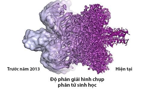 |
| New technique helps capture sharper images of biological molecules. Graphic: Guardian. |
Dubochet, professor emeritus at the University of Lausanne in Switzerland, developed a vitrification technique that allows biomolecules to freeze while maintaining their shape. Dubochet added water to the electron microscope. The liquid water evaporates in the electron microscope’s vacuum chamber, causing the biomolecules to crumble.
In the early 1980s, Dubochet achieved success with vitrifying water. He cooled water so rapidly that it solidified in liquid form around a biological sample, allowing the biomolecules to maintain their natural shape even in a vacuum.
The work of the three laureates “brings biochemistry into a new era,” providing innovative ways to observe the complex workings inside human cells at a resolution never before seen. With new imaging techniques, scientists can visualize everything from proteins that cause antibiotic resistance to the surface of the Zika virus.
Last year's Nobel Prize also went to microscopic research by scientists who created molecular machines, including the world's first molecular motor.
According to VNE
| RELATED NEWS |
|---|