Horrified by nearly 300 stones in patient's shoulder joint
Doctors at Saint Paul Hospital (Hanoi) recently performed arthroscopic surgery on a patient's shoulder and removed about 300 white stones that looked like pearls.
The patient is Mr. Nguyen Dong Toi, 37 years old, living in Dong Anh (Hanoi). According to the patient, the symptoms of left shoulder pain began to appear 6 years ago, the pain increased during daily activities, especially when moving the left arm. Recently, he felt his left shoulder joint stiffen, and then could not move. In addition to treatment with Western medicine, he also treated with oriental medicine methods such as taking medicine, applying hot compresses, massage, and acupressure, but the symptoms continued to get worse.
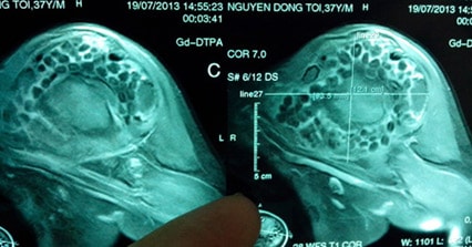 The image shows that the stones cover the entire left shoulder joint of patient Toi.
When he saw that his left shoulder joint was completely stiff, Mr. Toi went to Xanh Pon Hospital for examination, X-ray and MRI of his shoulder joint. After consulting with Dr. Nguyen Hanh Quang (Head of Orthopedic Trauma Department) and imaging diagnostics experts, the doctors concluded that Mr. Toi had synovial osteochondroma, and that there were many stones in his shoulder joint socket, which caused the symptoms of pain and reduced mobility. If he did not have timely surgery, there was a risk of losing the recovery of this joint.
Doctor Vuong Trung Kien, Deputy Head of the Orthopedic Trauma Department, who directly performed the endoscopic surgery on Mr. Toi, said it took 3 hours to remove nearly 300 stones, each with an average diameter of about 10mm, round and white like pearls. Although the disease was in a late stage, Mr. Toi was lucky to be discovered and operated on. After surgery, the patient recovered his shoulder joint movement very well and the pain symptoms gradually disappeared, but he still had to do rehabilitation for a while.
According to doctors, the synovial membrane is the innermost membrane of the joint capsule, which secretes synovial fluid to lubricate the joint's working surface. Synovial chondroma is an abnormal development of the cartilage structure in the synovial membrane, accompanied by calcium deposits. The calcium deposits grow larger, stick to the synovial membrane by a stalk and form clusters like bunches of grapes. In the late stages of the disease, "grapes" appear and fall into the joint, causing friction, compressing the cartilage, damaging the joint surface, degenerating the joint, and destroying the joint.
The disease only manifests in one joint, commonly the hip, knee, or shoulder. Diagnosis is mainly based on ultrasound, X-ray, CT scan, and MRI to determine and evaluate the stage of damage, thereby providing appropriate treatment.
When there is no phenomenon of calcium particles falling into the joint cavity, the patient can be treated with painkillers to reduce symptoms. On the contrary, when there are stones in the joint cavity, surgery to remove the stones is necessary, all other treatment methods are no longer effective. Previously, doctors used the open surgery method to cut the damaged synovial membrane, and at the same time remove the stones in the joint cavity. This method has the limitation of difficult stone removal, large incision and cutting the synovial membrane causes damage to the joint, so the surgical results are quite limited.
Nowadays, thanks to the endoscopic surgery method, stone removal becomes easier, avoiding the risk of remaining stones, and at the same time the synovial membrane is preserved, so the ability to recover the joint is very good. The endoscopic surgery method also allows for subsequent surgeries when the patient has recurrent stones in the joint cavity.
According to VnExpress - TL


