Surgery to remove 45kg tumor for young man who had to crawl on the street
The tumor on S.'s body kept getting bigger, taking up his entire back and buttocks, making it impossible for him to walk normally, almost forcing him to crawl on the road.
The tumor occupied the entire back of the buttocks and almost the entire thigh to the left calf. The tumor was so large that the patient could not walk normally. To move, S. almost had to crawl on the street or use a wheelchair because he did not have enough strength to carry the tumor.
The family said S. was the second child in the family. As he grew older, the tumor on his back and thighs grew larger, his legs swelled to the size of an elephant's, but the family was poor and did not have money for surgery. There was a time when the tumor was so large that it burst, causing the patient to bleed to the point of fainting. After that, the family borrowed money from everywhere to take their child to the doctor.
|
| Huge tumor on the patient's back, buttocks down to his legs. |
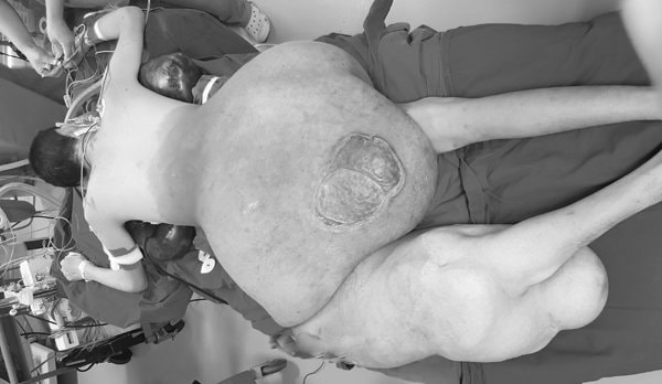
According to Associate Professor Ha, when arriving at Viet Duc Hospital, the patient was in a state of severe anemia, his body exhausted due to chronic blood loss. The tumor ruptured, leaving a deep ulcer larger than a hand that has not healed for years. Due to having to carry the large tumor for too long, S's left femur neck was also deformed and broken.
Difficult surgery 3, transfused 5 liters of blood
Associate Professor Ha diagnosed the patient with neurofibromatosis. However, when ordering an MRI to differentiate the tumor from the nerves, there was a problem because the tumor was too large to be placed in the imaging cage.
This is very dangerous because during tumor removal it is easy to damage the sciatic nerve, leading to paralysis of the lower limbs.
To engrave, the ultrasound machine must be brought to the operating room and the path of the nerve must be examined as the surgery progresses.
The second difficulty is that the tumor is too large, if you try to remove the entire tumor in one surgery, it may cause the patient to die on the operating table. The optimal solution is to perform surgery in multiple stages, at least twice.
The first surgery will remove as much of the tumor as possible from the patient’s back and buttocks. If the patient recovers after the first surgery, the tumor will be removed from the left thigh and knee, and the hip joint may even have to be removed if there is too much bleeding.
The third difficulty was that the tumor had a lot of blood vessels, which increased the risk of severe bleeding during surgery. Therefore, the doctors had to use two large ultrasonic knives, which were only used to stop bleeding during liver resection, to operate and seal the blood vessels at the same time, reducing the risk of bleeding.
Identifying this as a complicated surgery, the Hospital Board of Directors organized a consultation with a series of leading experts from nearly 10 specialties: Hematology, blood transfusion, kidney dialysis, diagnostic imaging, anesthesia, orthopedics, etc. in coordination with doctors from the Department of Plastic Surgery.
|
| Part of the tumor was removed. |
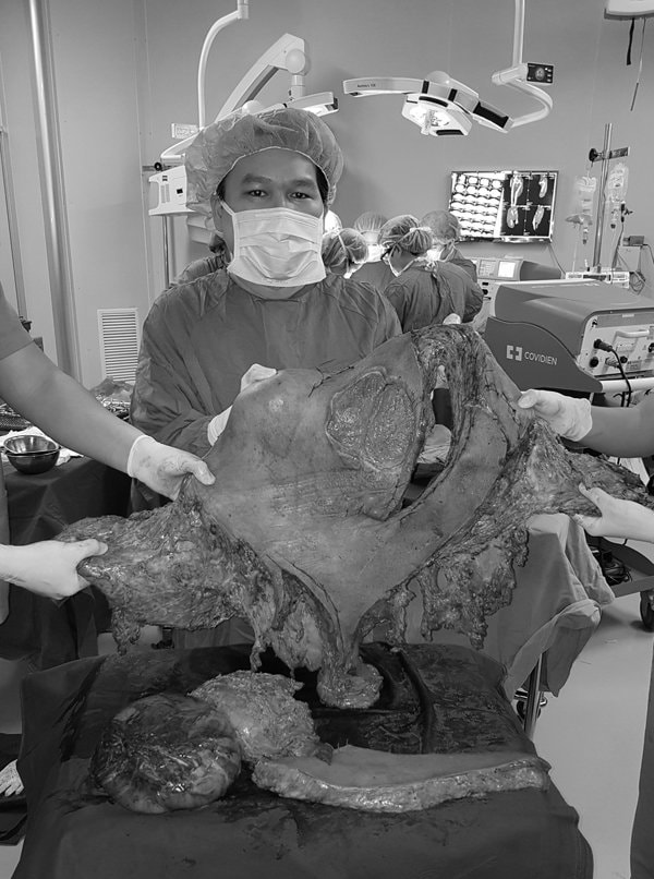
The surgical team mobilized more than 10 surgeons divided into 3 groups and 20 staff, doctors during surgery and anesthesia. After more than 8 hours of surgery, the team removed the entire tumor in the back and buttocks with a weight of about 23kg. The total amount of blood that had to be transfused to the patient was more than 5 liters.
Up to now, more than 1 month after surgery, the patient's health has improved, passed the intensive care phase but still has to closely monitor multiple organ function, prevent infection, and continue blood and protein transfusions.
It is expected that after 3-6 months, if the patient recovers well, another surgery will be performed to remove the tumor in the left thigh and knee.
Neurofibromatosis is caused by a genetic mutation on chromosome 17, most commonly manifesting on the skin. According to world medical literature, the genetic rate of this disease is 50/50.
The first symptoms are detected from a young age such as cafe au lait spots on the skin, freckles often found in the armpits or groin, tumors in the eyelid area, large neurofibromas on half of the face, head, neck (horse mane), upper limbs (bear paws), lower limbs (elephant feet) or giant tumors on the entire back and buttocks of the patient.
In addition, tumors can also be found in other locations such as the brain, adrenal gland tumors, enlarged liver, optic nerve tumors, and bones (can cause shortening or bone defects).
Diagnosis is mainly based on clinical picture and family history. Some variants of neurofibromatosis develop malignantly, accounting for 8-12% of cases.
Neurofibromas are very likely to progress to cancer, so the tumors must be regularly examined for pathology.


