New methods in diagnosing ovarian cancer
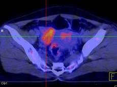
Many studies have shown that primary and metastatic ovarian cancer tissues strongly absorb FDG. Therefore, PET/CT imaging has provided information for ovarian cancer treatment with high sensitivity and efficiency.
According to 2003 statistics, in the United States there were approximately 25,400 new cases of ovarian cancer. Of these, approximately 14,300 died from ovarian cancer. Approximately 1.7% of women have a lifetime risk of developing ovarian cancer and 1 in 60 women will die.
The most common age is 70 - 74 years old, accounting for the highest rate among all age groups (57/100,000 people). Thanks to advances in surgery, chemotherapy and care services, the 5-year survival rate has increased over the decades, 37% in 1976, 41% in 1985, and 53% in 1998. In Hanoi, from 2001 - 2004, the incidence rate was 4.7/100,000 people, in Ho Chi Minh City it was 3.8%.

Diagnosis of ovarian cancer often requires a combination of different methods such as ultrasound, CT, MRI... Recently, FDG PET/CT imaging technique has been applied to diagnose ovarian cancer.
Many studies have shown that primary and metastatic ovarian cancer tissues strongly absorb FDG. Therefore, PET/CT imaging has provided diagnostic information, detected recurrence, metastasis, evaluated the effectiveness of ovarian cancer treatment... with high sensitivity and specificity. Ovarian cancer accounts for about 4% of diagnosed cancers and 5% of cancer deaths. Ovarian cancer often has a poor prognosis and is the main cause of death in adnexal cancers.
In the female reproductive system, FDG-PET imaging is mainly used in the initial diagnosis of cervical and ovarian cancer, as well as in the detection of recurrent lesions, monitoring treatment response, and assessing individual prognosis. In recent years, many studies have focused on evaluating the diagnostic value of FDG-PET/CT for ovarian and cervical tumors.
In Vietnam, PET/CT scans detect many patients with tumors in the ovaries and uterus. In addition, these patients often have increased FDG uptake on PET/CT images. The results of FDG-PET imaging are very valuable in detecting local and recurrent lesions and re-evaluating the stage.
Associate Professor, Dr. Mai Trong Khoa - Center for Nuclear Medicine and Oncology, Bach Mai Hospital.
According to (Kienthuc.net.vn) - M.D


