Malignant tumor in 2,000-year-old Egyptian mummy
Modern CT scanning technology allows doctors to detect a malignant tumor in the leg of a 2,000-year-old Egyptian mummy.
Doctors performed a full CT scan on the 2,000-year-old mummy found in an Egyptian tomb of a man nicknamed "Hen" at Crouse Hospital in Syracuse, New York, USA, Newsweek reported on December 13.
"The man had a tumor in his fibula, one of the two bones in his lower leg. It had all the hallmarks of a rare malignant tumor. It was a rare case of great interest," said Dr. Mark Levinsohn of Crouse University.
While the team of doctors cannot be certain the man died of cancer, the mummy has helped researchers better understand the disease that still affects people today.
This is not the first time Hen has been scanned. According to Dr. Levinsohn, he first scanned the mummy in 2006 in an attempt to diagnose the disease, but was unsuccessful.
"Over the past 10 years, the equipment has been upgraded. Back then we only had a 16-probe scanner, now we use a 320-probe scanner and can collect much more detailed information when scanning mummies," said Dr. Levinsohn.
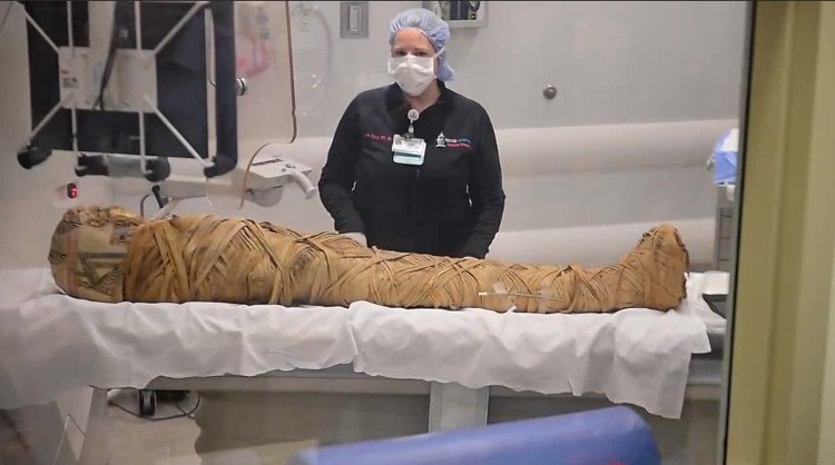 |
| Take the mummy to CT scan. |
The research team will continue tests on the mummy for another 2-3 months in the hope of finding more information about the man's cause of death.
According to Khoahoc.tv
| RELATED NEWS |
|---|


.jpg "Tiến sĩ, bác sĩ Nguyễn Khánh Toàn - người được bệnh nhân ung thư gửi gắm tin yêu")


.jpg "Đề nghị chấm dứt hoạt động cơ sở 'chữa ung thư bằng nước lọc ion kiềm' tại Quỳnh Lưu")